
MRI Small and Medium Joint — Wrist, Elbow or Ankle (MR zápěstí / loket / hlezno)
Type
Joint MRI
Duration
30 min
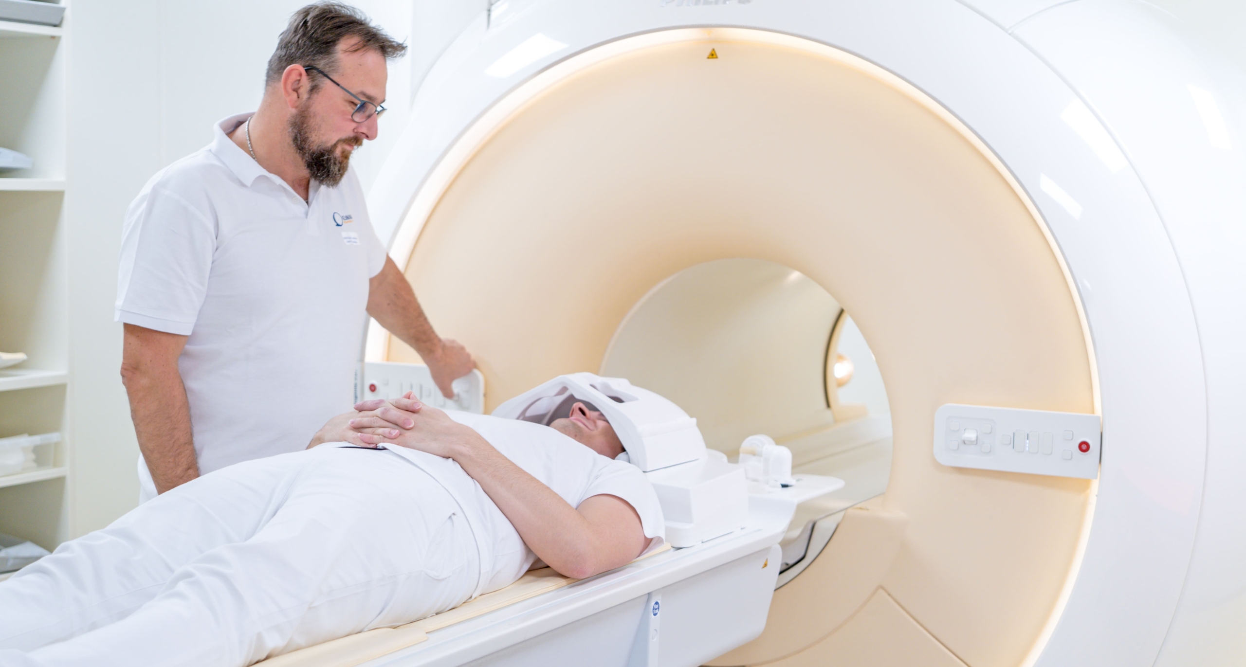
Klinika JL offers 3 Tesla MRI of the wrist, elbow or ankle — three commonly injured joints where soft-tissue detail is essential for surgical planning. The Philips 3 Tesla scanner resolves the fine ligaments of the wrist (TFCC, scapholunate and lunotriquetral), the lateral and medial elbow compartments, and the ankle lateral and medial ligament complexes and tendons. Each booking covers one joint at 9,000 Kč. The scanner bore accommodates studies in natural limb position; the wide-bore 70 cm Ingenia option provides additional patient comfort when joint positioning requires an unusual arm posture.
MRI of the wrist, elbow and ankle requires high spatial resolution to resolve the small, tightly packed structures in each joint. At 3 Tesla with a dedicated extremity coil, slice thickness can be reduced to 2–3 mm and in-plane resolution is sufficient to characterise individual ligament fibre bundles and focal tendon tears — detail that is often impossible at 1.5 Tesla. At the wrist, the triangular fibrocartilage complex (TFCC) is the primary structure of interest: tears are classified by the Palmar system (traumatic versus degenerative, peripheral versus central) and influence the choice between arthroscopic debridement and repair. The scapholunate and lunotriquetral interosseous ligaments are assessed in coronal plane at thin slices; partial tears are a common cause of chronic wrist pain in young adults after a fall on an outstretched hand. Tendon pathology — de Quervain tenosynovitis, flexor carpi ulnaris tendinopathy — is shown on axial sequences. Bone marrow oedema indicating occult fracture of the scaphoid or other carpal bones is seen on fat-saturated T2 or STIR before it is visible on X-ray. At the elbow, lateral epicondylosis (common extensor tendon origin) and medial epicondylosis are quantified on coronal T1 and PD images. Lateral ulnar collateral ligament tears are the MRI correlate of posterolateral rotatory instability; the UCL (ulnar collateral ligament) on the medial side is assessed in throwing athletes for partial or complete tear requiring Tommy John reconstruction. Osteochondritis dissecans of the capitellum is identified on coronal fat-saturated sequences. At the ankle, the lateral ligament complex (ATFL, CFL, PTTFL), the medial deltoid ligament, the Achilles tendon, peroneal tendons, posterior tibial tendon and spring ligament are all assessable. The anterolateral and posterior ankle impingement syndromes — common causes of chronic ankle pain after sprains — are shown on axial and sagittal sequences. Talar osteochondral lesions and sinus tarsi syndrome can be confidently characterised at 3 Tesla. Each booking covers one joint; patients needing two joints (e.g., bilateral wrists for carpal tunnel comparison) require two separate appointments. No contrast is typically needed for most sports-related ligament and tendon studies; gadolinium is reserved for tumour or infection suspected cases. The exam takes 20–40 minutes and requires no special preparation beyond removing metallic items and providing implant documentation.
Key Details
- Field strength
- 3 Tesla (Philips)
- Joints covered
- Wrist, elbow or ankle (one per booking)
- Key findings
- TFCC, ligaments, tendons, occult fractures
- Price
- 9,000 Kč per joint
Who Is This For?
TFCC tears, scapholunate injury, lateral epicondylosis, UCL tears in athletes, ankle ligament injuries, osteochondral lesions, occult fractures
What's Included
Preparation Required
Remove all jewellery and metal items from the hand, wrist, arm or ankle being examined. If you have surgical hardware in the joint, bring implant documentation. No fasting required. Bring your orthopaedic referral and any prior imaging of the joint.
9,000 Kč per single joint for self-pay patients (900,000 minor units). Price covers one joint per booking — wrist, elbow or ankle. Contrast adds 2,000 Kč if required. Covered by all major Czech health insurers on an orthopaedic referral.
- Category
- Diagnostic
- Duration
- 30 min