
Type
Abdomen MRI
Duration
35 min
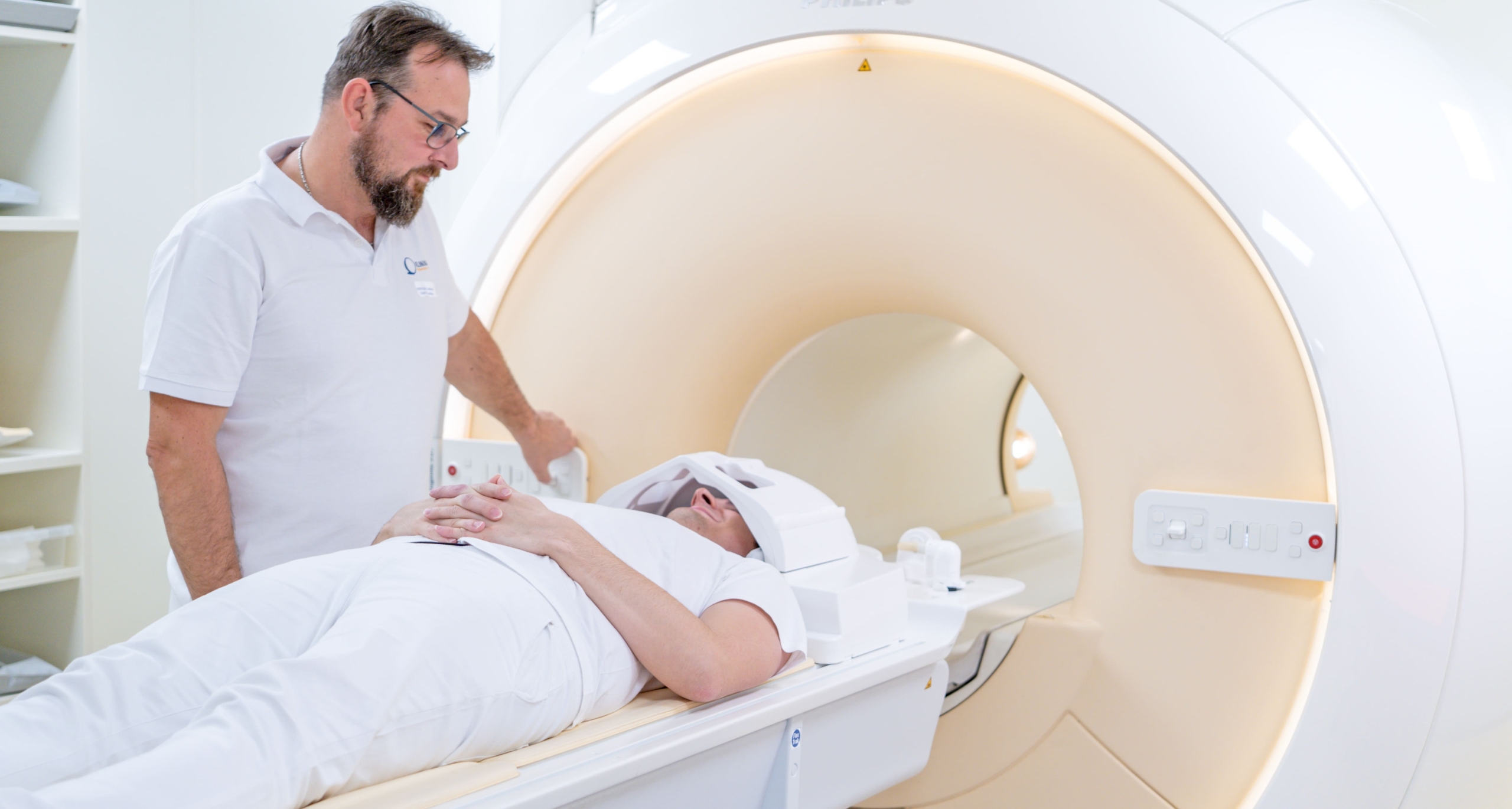
Liver MRI at Klinika JL uses the Philips 3 Tesla scanner with gadolinium contrast to provide definitive characterisation of liver lesions — distinguishing benign haemangiomas, focal nodular hyperplasia and adenomas from hepatocellular carcinoma and metastases in a way that ultrasound and CT cannot reliably achieve. Dynamic contrast sequences capture arterial, portal venous and delayed phases of enhancement, which are the hallmark pattern that identifies HCC. 3 Tesla field strength enables diffusion-weighted imaging (DWI) that adds functional information about cell density. Self-pay patients pay 9,000 Kč plus contrast. Three-to-four hour fasting before the scan reduces bowel motion artefact.
MRI is the gold-standard imaging modality for liver lesion characterisation, superior to CT for differentiating HCC from other focal liver lesions in the setting of cirrhosis and for detecting small metastases in colorectal cancer follow-up. At Klinika JL the Philips 3 Tesla scanner performs a multi-phase dynamic study: unenhanced T1 and T2 sequences establish baseline, then gadolinium is injected intravenously and the liver is imaged in the arterial phase (20–30 seconds after injection), portal venous phase (~60 seconds), and delayed or equilibrium phase (3–5 minutes). The pattern of enhancement across these phases identifies lesion type: HCC classically shows arterial hyperenhancement and portal venous washout (the LI-RADS criteria); haemangiomas show peripheral nodular early enhancement with centripetal fill-in; FNH shows central scar enhancement on delayed phase. Diffusion-weighted imaging (DWI) acquired at multiple b-values adds functional information: restricted diffusion (high signal on high-b-value images, low ADC value) indicates hypercellular tissue — malignant or inflammatory — and is used to detect small lesions below the resolution of structural sequences and to characterise large lesions. Four-year follow-up protocols for cirrhotic patients (biannual ultrasound, annual or biannual MRI) rely on the reproducibility of measurements at the same field strength; the clinic's Philips 3 Tesla system provides a stable reference for interval comparisons. Practical preparation: fasting for 3–4 hours reduces gastric and bowel motion that would blur the abdominal images; clear water is permitted. Breath-hold sequences require brief apnoea (8–15 seconds) multiple times during the scan; the radiographer will coach you through this via the intercom. The total scan time is approximately 35–45 minutes. Gadolinium contrast is contraindicated in severe renal insufficiency (eGFR <30 mL/min) — patients should mention any kidney disease history at booking. Results are reported by a qualified radiologist using standardised liver reporting systems where applicable (LI-RADS for cirrhotic patients). Digital images are available free via ePACS and Redimed; written reports are mailed to the referring doctor within 14 days.
Key Details
- Field strength
- 3 Tesla (Philips)
- Contrast
- Gadolinium IV, multi-phase dynamic (required)
- Preparation
- 3–4 hours fasting before the scan
- DWI
- Included — adds functional characterisation
Who Is This For?
Liver lesion characterisation, HCC staging in cirrhosis, colorectal cancer liver metastases, haemangioma vs adenoma vs FNH differentiation, hepatology follow-up
What's Included
Preparation Required
Fast for 3–4 hours before the exam (clear water is allowed). Remove all metal jewellery and items. Bring your referral and any prior abdominal imaging (CT, ultrasound). Inform staff of any kidney disease — eGFR needed if renal insufficiency is known. Gadolinium is contraindicated with severe renal disease. Drink extra fluids after the scan to clear the contrast agent.
9,000 Kč per scan for self-pay patients (900,000 minor units), plus 2,000 Kč for gadolinium contrast (required for liver characterisation). Total typical self-pay: 11,000 Kč. Covered by all major Czech health insurers on a gastroenterology or hepatology referral. Requires 3–4 hours fasting before the exam.
- Category
- Diagnostic
- Duration
- 35 min