
Type
MRI Scan
Duration
45 min
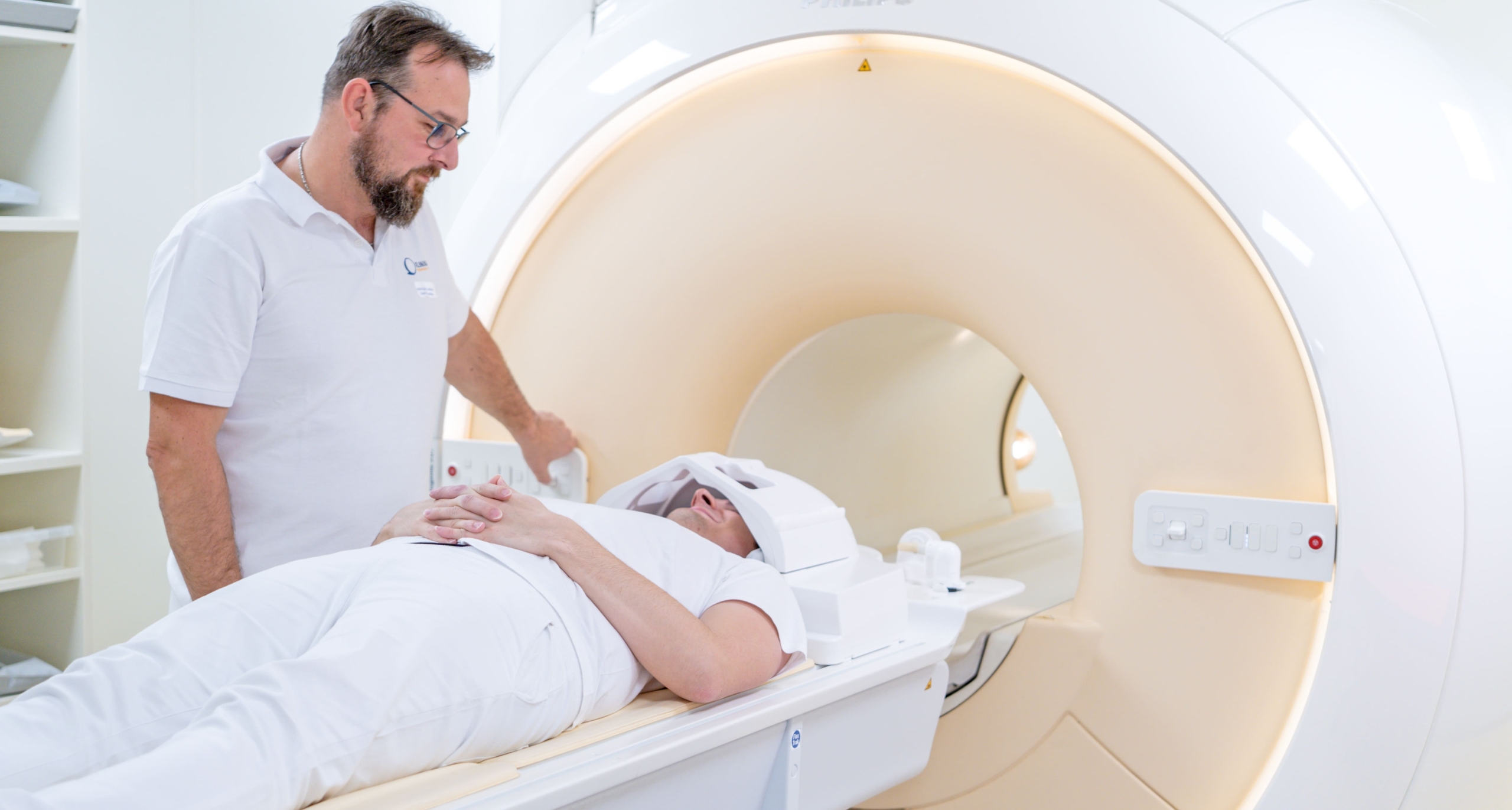
Fetal MRI (MR plodu) at Klinika JL is a specialised obstetric imaging study performed after 12 weeks gestation to evaluate fetal anatomy in cases where ultrasound is inconclusive — most commonly for central nervous system anomalies (ventriculomegaly, corpus callosum agenesis, cortical malformations), lung masses, diaphragmatic hernia and abdominal organ anomalies. At 15,000 Kč self-pay it is the most complex and highest-priced study on the clinic's list. No contrast is used. The Philips 3 Tesla scanner acquires rapid sequences designed to freeze fetal motion. The clinic requires a full bladder at the time of scan and a referral from a maternal-fetal medicine specialist or obstetrician.
Fetal MRI (MR plodu) at Klinika JL is the clinic's most specialised and highest-priced examination at 15,000 Kč, reflecting the complexity of the protocol, the need for rapid acquisitions to cope with fetal movement, and the dedicated time required for reporting fetal anatomy in up to 20 organ systems. The study is performed from 12 weeks gestation onwards — not in the first trimester — and without gadolinium contrast. The primary indication worldwide for fetal MRI is central nervous system anomalies detected or suspected on ultrasound. MRI exceeds ultrasound for brain sulcation and gyration assessment (cortical development maturity), corpus callosum and posterior fossa evaluation (Blake's pouch cyst vs Dandy-Walker vs mega cisterna magna distinction), ventriculomegaly grading and aqueductal stenosis, and periventricular and cortical heterotopias. These findings directly affect prognosis counselling and parental decision-making. Other major indications include: congenital lung lesions (CPAM/CCAM vs bronchopulmonary sequestration), diaphragmatic hernia (lung volume measurement on MRI predicts postnatal survival better than O/E LHR on ultrasound alone), abdominal wall defects (gastroschisis vs exomphalos, liver position), renal anomalies (bilateral renal agenesis, MCDK, duplex kidney complications), sacrococcygeal teratoma extent, and placenta and cord anatomy when ultrasound is inadequate. Fetal MRI technique: sequences are chosen to minimise scan time per image to freeze fetal motion. Single-shot T2-weighted sequences (SSFSE/HASTE) take 200–800 ms per slice and are the workhorse for brain and body; they are repeated in axial, coronal and sagittal planes relative to the fetal anatomy rather than the maternal body. EPI-based DWI can contribute to brain assessment. Gradient echo T1 is used for liver, bowel meconium (normally T1-hyperintense in the third trimester; abnormal when absent, suggesting bowel obstruction) and pulmonary assessment. Fetal movement — particularly in the second trimester — is the main image quality challenge. The radiographer monitors the scan continuously and repeats sequences in real time as needed. Breath-holding by the mother during individual sequences (8–15 seconds) eliminates maternal breathing motion; the fetus continues to move independently. The scan takes approximately 30–60 minutes depending on fetal position and cooperation. A full bladder aids lower uterine and placental visualisation when relevant. No preparation beyond normal hydration is needed. Reporting requires radiologists with dedicated fetal MRI experience; at Klinika JL reports follow obstetric conventions with fetal measurements calibrated to gestational age norms. The report is sent to the referring maternal-fetal medicine specialist for multidisciplinary team review.
Key Details
- Gestational age
- After 12 weeks only
- Contrast
- None — no gadolinium in pregnancy
- Primary indication
- CNS anomalies, lung lesions, diaphragmatic hernia
- Duration
- 30–60 minutes depending on fetal position
Who Is This For?
Ventriculomegaly, corpus callosum agenesis, cortical malformations, posterior fossa anomalies, congenital lung masses, diaphragmatic hernia lung volume, sacrococcygeal teratoma, bowel obstruction assessment
What's Included
Preparation Required
Drink approximately 500 mL water 1 hour before the scan — a comfortably full bladder improves lower uterine visualisation. Come prepared to lie still and practise brief breath-holds (8–15 seconds) during the scan. Bring your referral from a maternal-fetal medicine specialist or obstetrician with the documented gestational age and sonographic findings. No gadolinium will be used. This study is not performed in the first trimester (before 12 weeks).
15,000 Kč per scan for self-pay patients (1,500,000 minor units) — the highest-priced examination at this clinic, reflecting the specialised protocol and dedicated reporting time. No contrast agent used. Covered by all major Czech health insurers on a maternal-fetal medicine or obstetric referral. Not performed before 12 weeks gestation.
- Category
- Diagnostic
- Duration
- 45 min