

Type
MRI Scan
Duration
35 min
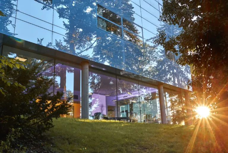
Brain MRI at Klinika Dr. Pírka is performed on a Siemens Magnetom Sola 1.5T scanner in the hospital's purpose-built imaging pavilion, opened May 2023. The department is led by MUDr. Zuzana Ryznarová, Ph.D., who supervises all head and neck imaging protocols. A brain MRI at 1.5T produces detailed, high-contrast images of brain parenchyma, white matter tracts, ventricular system, cerebellum, and brainstem, and is the standard investigation for headaches, neurological symptoms, suspected stroke, dizziness, or memory concerns. The clinic serves both self-pay patients and those referred by neurologists or GPs, with priority scheduling available for private patients. Being embedded within a full multi-specialty hospital means neurology and orthopaedic consultants are available on the same site to review findings and plan next steps without the patient needing to travel.
Klinika Dr. Pírka operates brain MRI on its Siemens Magnetom Sola 1.5T system in the dedicated imaging pavilion built in 2023. The Sola platform's 70 cm bore is wider than older closed-bore 1.5T designs, which meaningfully reduces the feeling of confinement that some patients associate with brain scanning — the head is positioned at the isocentre of the bore but the body remains largely outside the narrowest section, which most patients tolerate well. Brain MRI at 1.5T provides diagnostic-quality imaging for the vast majority of clinical indications including: headache workup (identifying structural causes such as tumours, vascular malformations, or white-matter lesions), stroke and TIA assessment (DWI and FLAIR sequences for acute ischaemic lesions), multiple sclerosis surveillance (lesion load, new activity), epilepsy evaluation (hippocampal volume, focal dysplasia), vertigo and balance disorders (posterior fossa, inner ear canals, cranial nerve VII/VIII), dementia assessment (cortical atrophy patterns, hippocampal atrophy grading), and screening in patients with a family history of intracranial aneurysm. The radiologist can add gadolinium contrast enhancement when enhancement patterns are needed (e.g., leptomeningeal disease, blood-brain barrier breakdown, tumour vascularity), subject to contraindication screening and renal function check. At Klinika Dr. Pírka, the neurology department — staffed by MUDr. Věra Najmonová (EMG specialist) and MUDr. Jana Horáková — can refer patients directly to the in-house MRI department for expedited scanning and same-campus reporting. This closed-loop pathway is particularly efficient for neurology outpatients who have their initial consultation at the clinic and can have imaging scheduled the same or next working day rather than being redirected to a separate radiology centre. Preparation for brain MRI is straightforward: no fasting is required for a non-contrast scan. Gadolinium contrast is given intravenously if indicated; for this you should fast for 4 hours beforehand and the team will screen for any history of allergic reactions to contrast agents and check renal function. Standard MRI safety rules apply — remove all metallic objects (earrings, hairpins, hearing aids). Inform the booking team at the time of scheduling about any metal implants in the head or neck region (dental implants embedded in bone are generally safe but braces, bridges, and certain older surgical clips are not), any cochlear implant, or any electronic implanted device. Brain MRI scans at 1.5T typically take 25–40 minutes depending on the number of sequences requested. Standard protocols include T1-weighted, T2-weighted, FLAIR, and diffusion-weighted imaging (DWI). Additional sequences such as SWI (susceptibility-weighted for micro-haemorrhages), MRA (magnetic resonance angiography for vessel assessment), or spectroscopy may be added when clinically indicated, extending the session slightly. After the scan, MUDr. Ryznarová prepares a written report which is included in the self-pay fee. Reports are typically available within a few working days and can be forwarded to your referring physician by email. Digital images are provided on CD or in digital format on request. Self-pay bookings are made via the dedicated MRI line (+420 326 375 470, Mon–Fri 9:00–15:00) or by email to [email protected].
Key Details
- Scanner
- Siemens Magnetom Sola 1.5T — 70 cm bore
- Protocols
- T1, T2, FLAIR, DWI; gadolinium contrast available
- On-site neurology
- Same-campus neurologist consultations available
- Report
- Written radiologist report by MUDr. Ryznarová, Ph.D.
- Booking
- [email protected] or +420 326 375 470
Who Is This For?
Patients with persistent headaches, neurological symptoms, suspected stroke history, MS monitoring, epilepsy workup, memory concerns, vertigo, or family history of intracranial aneurysm
What's Included
Preparation Required
No fasting required for non-contrast brain MRI. For gadolinium contrast (if requested by your physician): fast for 4 hours beforehand. Remove all metallic items — earrings, hairpins, hearing aids. Inform the team about any head or neck implants, cochlear implants, or electronic implanted devices. No recovery time after the scan — you can drive and resume all activities immediately.
Approx. 3 500 Kč per brain MRI examination (pricing on request — contact [email protected] or +420 326 375 470, Mon–Fri 9:00–15:00). Includes the scan on the Siemens Magnetom Sola 1.5T and a written radiologist report by MUDr. Zuzana Ryznarová, Ph.D. Czech public health insurance accepted with a valid neurology or GP referral; self-pay patients receive priority scheduling in the private MRI pavilion.
- Category
- Diagnostic
- Duration
- 35 min
"{"author":"Dalibor Jelínek","platform":"Firmy.cz","date":"April 2026","text":"Profesionálové každým coulem — professionals in every way. The care was..."