Type
General Skin Treatment
Duration
20 min
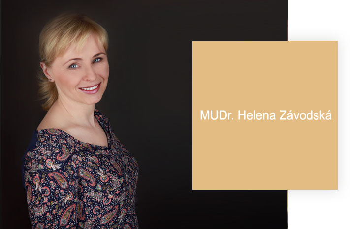
Removal of benign skin growths including skin tags, seborrheic keratoses, fibromas, dermal naevi and similar surface lesions using FOTONA 4D Er:YAG laser ablation or conventional surgical excision. MUDr. Helena Závodská determines the method based on lesion type, depth, size and location. Laser removal offers precise ablation with minimal surrounding damage and no sutures for superficial lesions; surgical excision is preferred for deeper or larger lesions and provides a specimen for histopathological analysis if indicated.
Benign skin growths — such as skin tags (acrochordons), seborrheic keratoses, dermatofibromas, syringomas, milia and small epidermal naevi — are extremely common and are driven by ageing, genetics, skin friction and mild chronic irritation. While medically benign, they may be cosmetically bothersome, catch on clothing or jewellery, or require clinical assessment to confirm their benign nature before removal. At AE Derm MUDr. Helena Závodská assesses every growth clinically before deciding on removal method. A dermatoscopic examination is performed first: the dermoscope reveals the internal structure of the lesion, allowing distinction between truly benign growths and any that have features warranting diagnostic excision rather than simple ablation. Once confirmed benign, the removal method is chosen based on lesion characteristics. Laser ablation with the FOTONA Er:YAG 2940 nm mode offers very precise, layer-by-layer removal of the lesion's tissue with controlled depth. Er:YAG's high water absorption means that each pulse removes a precise thickness of tissue with minimal thermal spread to the surrounding skin; the operator can stop at exactly the right depth, leaving a clean wound base that heals with minimal scarring. This technique is particularly suitable for flat, superficial lesions such as seborrheic keratoses, small fibromas, skin tags on the face or neck and milia clusters. Local anaesthetic is injected before the procedure; the ablation itself takes only a few minutes. The wound is then treated with antiseptic and a small dressing; the healing crust separates within ten to fourteen days, leaving a temporary pink area that fades over two to four weeks. For lesions that require a tissue sample for histopathology — any pigmented lesion with atypical dermoscopic features, a growth whose clinical behaviour has changed, or a patient with a personal history of skin malignancy — surgical excision under local anaesthetic is performed with standard scalpel technique and closure with fine sutures. The specimen is sent for pathological examination. Sutures are removed after seven to ten days depending on location. Following any growth removal, sun protection is particularly important during the healing phase to prevent post-inflammatory pigmentation of the healing wound.
Key Details
- Methods available
- Er:YAG laser ablation or surgical excision
- Pre-removal assessment
- Dermoscopy to confirm benign nature
- Histopathology
- Surgical excision provides specimen if needed
- Healing
- Crust separates at 10–14 days; full resolution 2–4 weeks
Who Is This For?
Adults with cosmetically bothersome or irritating benign skin growths such as skin tags, seborrheic keratoses, small fibromas or milia who want physician assessment and precise removal
What's Included
Preparation Required
Arrive with clean skin. Inform the doctor of any blood-thinning medications (aspirin, ibuprofen, anticoagulants) — these may need to be paused. Do not apply make-up or self-tan to the area.
From approximately 2,000 Kč per growth (market reference — confirm at consultation as pricing depends on size, number and method). Contact [email protected] for an individual assessment.
- Category
- Skin Treatments
- Duration
- 20 min
"Always on time, no problems with appointments. The doctor is very professional and I felt confident throughout. — Josef Šoltes, Firmy.cz"