Type
General Skin Treatment
Duration
30 min
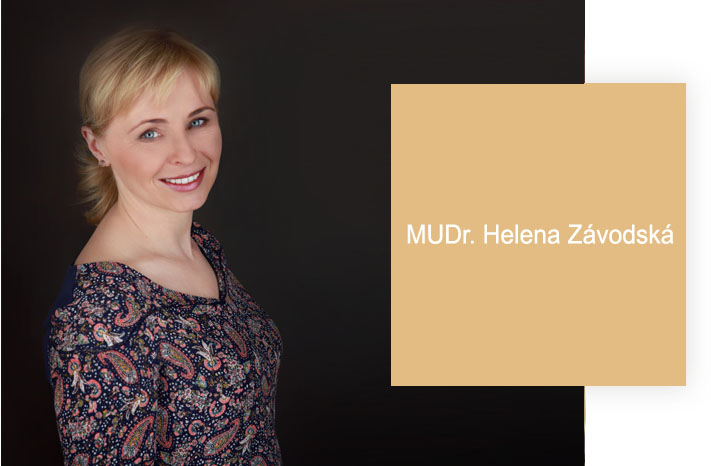
Digital dermoscopy examination of moles, pigmented lesions and other skin changes using a dermatoscope with archival software. MUDr. Helena Závodská is a trained dermatologist who performs ABCD criteria assessment — evaluating Asymmetry, Border, Colour and Diameter/Dermoscopic structures — with risk stratification software. Photo documentation is stored for comparative analysis at follow-up visits, enabling detection of changes over time that would not be visible to the naked eye.
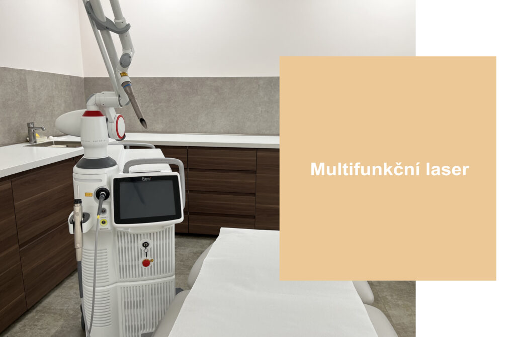
Dermoscopy (also called epiluminescence microscopy) is a non-invasive diagnostic technique that uses magnification and controlled illumination to visualise structures within the skin that are invisible to the unaided eye. A standard dermatoscope illuminates the skin surface while eliminating reflective glare, revealing pigment network patterns, vascular structures, regression areas and other dermoscopic criteria in melanocytic and non-melanocytic lesions. Digital dermoscopy takes this further: images are captured and stored electronically, linked to the anatomical location, and can be retrieved and compared at subsequent visits to detect any changes in size, colour, structure or pigment distribution. At AE Derm MUDr. Helena Závodská uses the digital dermatoscope with archival and comparative software. She applies the ABCD dermatoscopy rule systematically — Asymmetry of shape and structure, Border regularity and abruptness, Colour uniformity and number of distinct colours, and Dermoscopic structures including dots, globules, regression and vascular patterns. The software also performs automatic pattern recognition and provides a risk stratification score, which MUDr. Závodská reviews in clinical context with the patient's history. For patients with multiple moles, a whole-body or targeted mapping can be performed in which all lesions of concern are documented and indexed. At follow-up visits — typically every six to twelve months for surveillance patients — the same lesions are re-imaged and compared side by side, allowing detection of even subtle change that would not be perceptible without baseline photos. This time-comparative analysis is the primary advantage of digital over conventional dermoscopy: early change is detectable, enabling timely referral for excision if needed. The procedure is entirely painless and non-invasive, requiring no preparation beyond avoiding artificial tan products on the examination day. Patients who have had prior melanoma or who have many atypical moles, a family history of melanoma, significant cumulative sun exposure or previous sunburn, fair skin or red/blonde hair should consider annual surveillance. MUDr. Závodská also offers ongoing oncological dispensary care for patients with diagnosed skin malignancy who require specialist follow-up.
Key Details
- Equipment
- Digital dermatoscope with archival software
- Assessment
- ABCD criteria + pattern recognition risk scoring
- Time comparison
- Yes — images stored and compared visit to visit
- Invasiveness
- None — entirely non-invasive
Who Is This For?
Adults concerned about moles or skin changes; those with a history of melanoma, many atypical moles, significant sun exposure or family history of skin cancer; annual surveillance patients
What's Included
Preparation Required
Do not apply self-tan to examination areas for two weeks before. Arrive without body lotion or cream on the skin. Bring any previous dermoscopy reports or photos from other clinics if available.
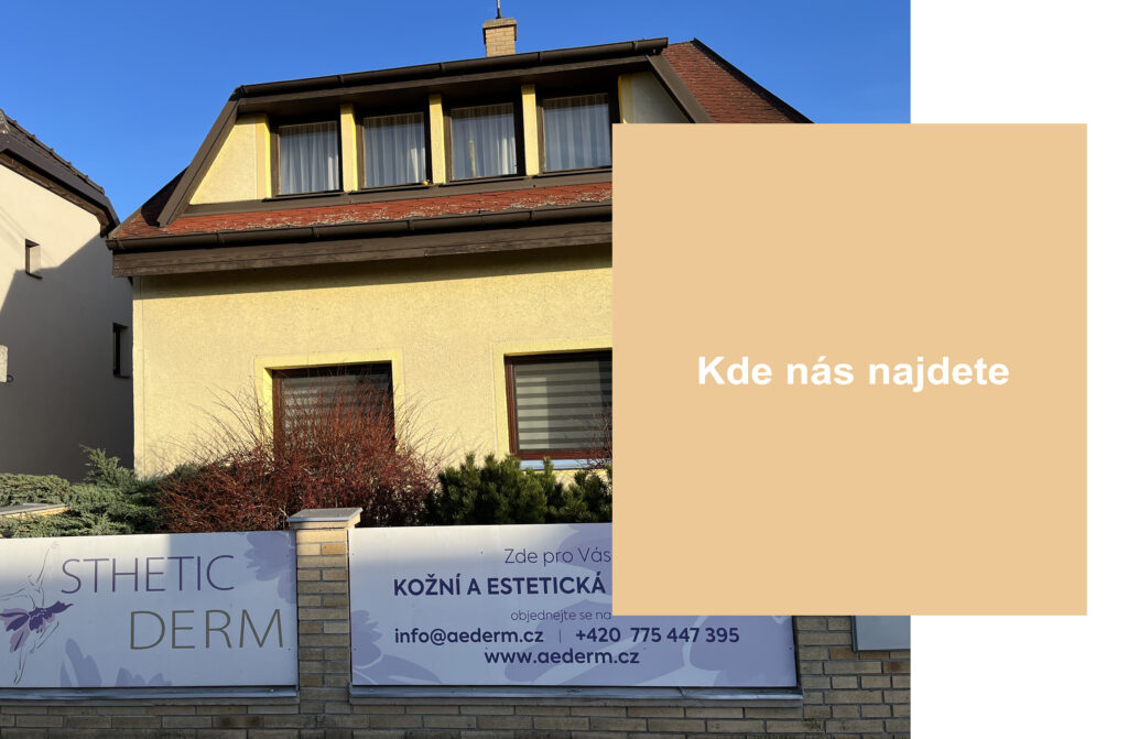
From approximately 1,000 Kč per examination (market reference for digital dermoscopy — confirm at consultation). Includes photo documentation and ABCD risk assessment. Contact [email protected] or call +420 775 447 395.
- Category
- Skin Treatments
- Duration
- 30 min
"Always on time, no problems with appointments. The doctor is very professional and I felt confident throughout. — Josef Šoltes, Firmy.cz"